
Kidney Stones
Show video transcript
Kidney stones are very common, they occur in approximately 12% of people.1
They are very common and unfortunately, extremely painful.
Patients usually present with pain in the loin which radiates down to the groin area and they often have nausea and vomiting.
It’s important to check that they don’t have a fever. One cause of fever could be an obstructed and infected kidney. This is a medical emergency and we have to drain that kidney straight away.
Our management generally depends on initially diagnosing the problem.
So the way we diagnose the problem is that we do
- A urine test: we check if there’s blood in the urine. That gives us an indication that there might be a kidney stone. This also allows us to check that there’s no infection.
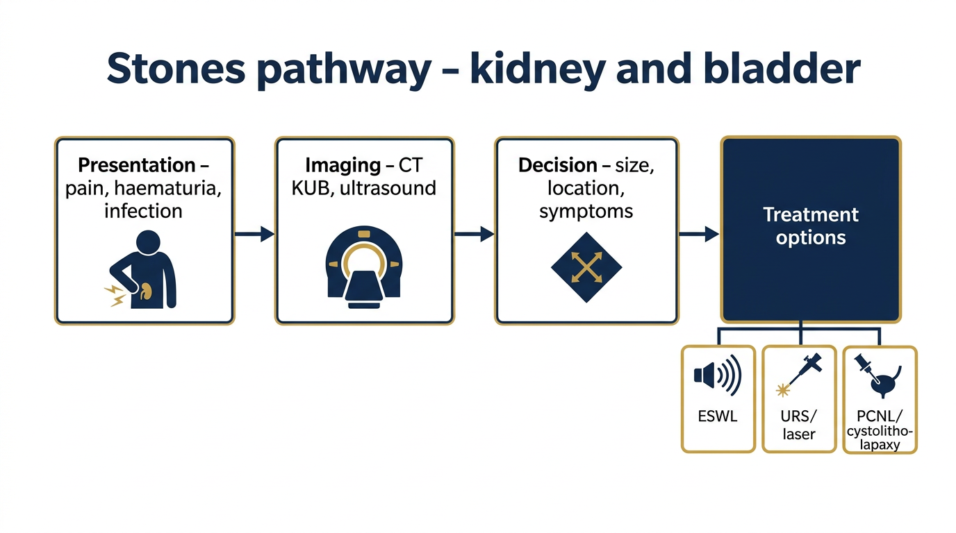
- Imaging tests: The imaging test that we do these days is a CT scan, a non-contrast spiral CT scan and this is very, very accurate2 and it usually tells us where the stone is, the size of the stone and the degree of blockage or obstruction to the system.
The management then usually depends on the patient’s symptoms, do they have ongoing pain? Do they have a fever? What is the size and location of the stone?
So if patients have a small stone, say less than 4mm we will monitor them conservatively, if the pain settles, we will monitor them, give them analgesia (pain relief). They’re usually discharged, we then do regular scans and wait a certain time period and let them see if they pass the stone.
If they haven’t passed the stone in a reasonable time period we’ll then intervene and remove that stone.
If they have a larger stone greater than 6mm the chance of that stone passing is very low3, so we would generally treat that stone.
The stone may be located in the ureter, the tube that comes from the kidney to the bladder and we generally treat that with ureteroscopy which I’ll explain shortly.
Sometimes we also find very large stones, stag horn stones which occupy a lot of the kidney and the drainage system. Stag horn stones are quite complex and they require different forms of treatment.
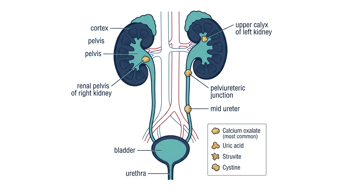
There are essentially four types of stones.4
CALCIUM STONES - The commonest stones are made of calcium - either calcium oxalate or calcium phosphate and hence you can see them on plain x-rays.
They comprise about 90% of all stones5
STRUVITE STONES - Sometimes you get infective stones or struvite stones which are caused by urea splitting bacteria and usually occur in young females who have recurrent urinary tract infections
URIC ACID STONES - The other type of stone are uric acid stones which are related to patients with gout who produce increased levels of uric acid.
CYSTEINE STONES - Very rarely patients will have cysteine stones, a congenital abnormality where they have abnormal absorption of cystine from the urine they form these stones.
KIDNEY STONES TREATMENT
In terms of the treatments that patients have, a ureteroscopy is generally used when the stone is in the ureter, that is the tube from the kidney to the bladder.
We pass a thin telescope either in through the eye of the penis in males or the urethra in females.
We go up the ureter which is the tube that joins the kidney to the bladder and we get to the stone.
Usually the stone is too big to come out by itself and we have to fragment the stone.
So we usually break the stone with either a LITHOCLAST6 which is an instrument that’s like a mini jackhammer or a HOLMIUM LASER7 which is very precise, it’s like a surgical knife, it brakes the stone in very precisely and then we take out all the little pieces with a basket, a DORMIA BASKET8 and remove the stone.
I usually plan to remove all the stone.
Some people break the stone and allow patients to flush the stones out but my aim is usually for complete clearance.
Now I also do ureteroscopy for complex stones in the kidney where we go up with a flexible ureterscope that bends all the way up to the kidney and we can go around the corner of the kidney and treat the stones and treat it with a laser.
So in patients that have had stones that are not suitable or lithotripsy or lithotripsy doesn’t work then we would use flexible ureteroscopy and laser treatment.
Usually after ureteroscopy we will put a stent in, a plastic tube that curls between the kidney and bladder which ensures that urine flows out and then we remove that either with local anaesthetic gel or a light anaesthetic in usually one to two weeks.
Now stones that are present in the kidney are usually treated with LITHOTRIPSY.
Lithotripsy is shockwave treatment that’s given via an external machine.
A patient lies down, usually under an anaesthetic, for about 40 minutes or so and has external “shockwave energy” given to the stone. Remarkably this stone can fragment and break into little pieces and then we wait for the patient to pass these little pieces.
Success rate for lithotripsy is about 80 to 85%.9
Some stones you don’t have success straight up and you have to have multiple treatments and some stones unfortunately are refractory to lithotripsy and if they are then we do the flexible cystoscopy/ureteroscopy and in suitable cases, laser treatment.
Sometimes if we have a very large stone in the kidney, so called stag horn stone we will do a percutaneous nephrolithotomy. This is keyhole surgery where we puncture the side of the kidney, we go into the kidney, we find the stone, we break the stone either with a lithoclast which is a mini jackhammer or with an ultrasound which breaks up some of the fragments or with a laser and then we remove the fragments that way.
The patients are usually in hospital for two days.
There is some concern with percutaneous nephrolithotomy because we do have to go through the kidney for that procedure10 but certainly it’s preferable to a big open operation.
Now occasionally I’ve also removed stones laproscopically with keyhole surgery.
So if we have a very large stone in the kidney, renal, pelvis or ureter we can go in with keyhole surgery and remove it and patients are just in hospital overnight.
The important thing with kidney stones is to get the stones analysed, get blood tests done and to see if patients have abnormal uric acid or calcium level and check that there’s no abnormality.
Sometimes we do a parathyroid level because the parathyroid gland here produces hormones that can lead to increased calcium levels.
If patients have recurrent stones I usually refer them to one of my colleagues, one of the renal physicians for a full metabolic evaluation so that we can try to minimise the chance of stone recurrence.
So with kidney stones is to ensure that you do whatever you can to prevent them recurring.
The most important thing is to increase fluid intake, it’s very simple, there’s little molecules in the urine and they’re close together, they join, they form a stone, when they’re far apart they don’t so you have to drink two to three litres a day.
There’s evidence that home made lemonade is better than water itself.11
I tell patients that they should ensure that their urine is clear and they’re passing urine every two to three hours.
So that’s really what we do with kidney stones.
It’s a common condition but there’s lots of treatment, a lot of good treatments available and if you have recurrent stones you should have a full metabolic evaluation to minimise the chance of them recurring.
At a glance
- What they are: Hard deposits of minerals and salts that form inside the kidney and can travel into the ureter, the tube that carries urine from the kidney to the bladder.1, 4
- How common: Around one in ten people will form a kidney stone during their lifetime, and after a first stone roughly half will form another within 5 to 10 years without preventive measures.1, 22
- How they present: Severe loin pain radiating into the groin, often with nausea and vomiting. Fever with stone pain is a medical emergency - see below.
- How they are diagnosed: A urine test plus a non-contrast CT of the urinary tract is the standard of care. Non-contrast CT is highly accurate for urinary stones, with pooled sensitivity around 95 per cent and specificity around 96 per cent reported in the EAU (European Association of Urology) 2025 guideline’s own systematic review.2, 22, 37
- What size means: Stones of about 4 mm or less often pass on their own (approximately 75 to 90 per cent, depending on exact size and location); stones larger than 6 to 7 mm are less likely to pass and usually need treatment.3, 22, 34
- Treatment options: A small stone with mild symptoms can be observed. Active treatments include medical expulsive therapy for selected ureteric stones, extracorporeal shockwave lithotripsy (ESWL), ureteroscopy with holmium laser lithotripsy (URS/RIRS), percutaneous nephrolithotomy (PCNL) for large or stag-horn stones, and occasionally laparoscopic or robotic stone surgery for selected complex cases.22, 23
- Prevention matters: After a first stone, lifestyle and dietary changes - especially increased fluid intake - reduce the chance of another stone, and targeted medical therapy is available for recurrent stone formers.22, 24, 33
What are kidney stones?
Kidney stones are solid deposits that form when minerals and salts in the urine become concentrated enough to crystallise. They can sit quietly inside the kidney for months or years, or they can move into the ureter and cause sudden severe pain - renal colic.
Most patients present with pain in the loin that radiates around the flank into the groin, often with nausea and vomiting.1 The pain is usually severe, comes in waves, and is unrelated to movement - unlike musculoskeletal pain, sitting still does not make it better.
Kidney stones are common: approximately one in ten people is affected during their lifetime, and after a first stone there is a meaningful chance of forming another one without preventive measures.1, 22 The exact risk of recurrence depends on the type of stone and the underlying cause, which is why identifying both is an important part of long-term management (see Preventing recurrence below).
When kidney stone pain is an emergency
Pain from a kidney stone together with a fever is a medical emergency. It suggests that the stone is obstructing the kidney and that urine upstream of the blockage has become infected - a condition called obstructive pyelonephritis or infected hydronephrosis. International guidelines recommend urgent decompression of the kidney (usually by placing a ureteric stent or a percutaneous nephrostomy tube), together with immediate intravenous antibiotics and, if necessary, intensive care support.22
Seek emergency care without delay if any of the following occurs:
- Loin or flank pain with a fever above 38 degrees Celsius, shaking chills, or rigors.
- Pain so severe that it cannot be controlled with ordinary analgesics.
- No urine output or blood with clots in the urine.
- Pain associated with confusion, fast heart rate, or feeling very unwell.
These features can indicate sepsis from an infected obstructed kidney and need hospital assessment immediately.22
How kidney stones are diagnosed
Assessment typically includes:
- Clinical examination and urine test. Blood in the urine is common with stones. A urinary infection needs to be excluded urgently, particularly if there is any fever.22
- Non-contrast CT of the urinary tract (CT KUB). This is the imaging study of choice. In the EAU 2025 guideline’s own systematic review it has pooled sensitivity around 95 per cent and specificity around 96 per cent for urinary stones, and it shows the size, number, location, and density of the stone or stones, as well as the degree of obstruction of the kidney above.2, 22, 37 Radiation exposure is minimised using a low-dose protocol wherever possible.
- Ultrasound is sometimes used as an initial test - particularly in pregnancy, in children, or when CT is not immediately available - and is sensitive for hydronephrosis (swelling of the kidney due to obstruction) but less accurate than CT for the stone itself.22
- Blood tests check kidney function, markers of infection, and - in patients with recurrent stones - calcium, uric acid, and sometimes parathyroid hormone. More detailed metabolic testing follows if indicated (see Preventing recurrence).
- Stone composition analysis (when a stone is passed or retrieved) directly identifies the type of stone and helps tailor prevention.22
Types of kidney stones
There are four main types of kidney stone.4, 5, 35
- Calcium stones. Around 80 per cent of stones are calcium stones, usually calcium oxalate and less commonly calcium phosphate.5, 22, 35 They are visible on plain x-rays and on CT.
- Uric acid stones. About 5 to 10 per cent of stones.5, 35 They are associated with gout, high dietary protein intake, obesity, and persistently acidic urine. Unlike calcium stones, pure uric acid stones do not show on plain x-rays and are less dense on CT.
- Struvite (infection) stones. Caused by urea-splitting bacteria, typically in women with recurrent urinary tract infections. They can grow rapidly into the branching shape of the kidney’s collecting system - a stag-horn stone - and often require more than one procedure to clear.22
- Cystine stones. Uncommon. Caused by an inherited defect in how the kidney reabsorbs the amino acid cystine, leading to very high cystine levels in the urine and recurrent stones from an early age.
Rarer stone types - drug-related stones, matrix stones, xanthine stones - also exist but together account for a very small minority of cases.22
How treatment is chosen
Modern guidelines recommend that treatment be individualised to four main factors: the patient’s symptoms, the size of the stone, its location, and the presence of any obstruction or infection.22, 23
Size
- Stones of about 4 mm or less. Approximately 75 to 90 per cent pass spontaneously with simple measures - adequate fluid intake and analgesia - depending on exact size and location, so observation is usually reasonable provided pain is controlled and there is no infection.3, 22, 34
- Stones 5 to 6 mm. Intermediate. Many will still pass; selected patients may be offered medical expulsive therapy (see below).
- Stones larger than 6 to 7 mm. Less likely to pass on their own. Active treatment is generally recommended.3, 22, 23, 34
- Stones larger than 2 cm, or stag-horn stones. Usually treated with percutaneous nephrolithotomy (PCNL) as first-line, in line with both EAU 2025 and AUA 2026 guideline recommendations (AUA 2026: Moderate Recommendation, Evidence Level Grade B; EAU 2025: supported by high-quality comparative evidence).22, 23
Location
- Stones in the ureter are usually treated with semi-rigid or flexible ureteroscopy and laser lithotripsy, with ESWL an alternative in selected cases.22, 23
- Stones in the kidney can be treated with ESWL, flexible ureteroscopy with laser lithotripsy (retrograde intra-renal surgery, RIRS), or PCNL depending on size and location inside the kidney.22, 23
Obstruction and infection
A stone that is obstructing the kidney, particularly with any sign of infection, is a surgical emergency - the kidney is drained first (stent or nephrostomy) and the stone is treated at a later date once the infection has cleared.22
Active surveillance for small, symptom-free kidney stones
This is an option in some cases. Selected small, non-obstructing kidney stones that are not causing symptoms can be kept under regular imaging review rather than treated immediately, in line with current international guidelines.22, 23
Treatment options
Urology NSW offers the full range of contemporary kidney stone treatments - medical expulsive therapy, ESWL, semi-rigid and flexible ureteroscopy with holmium laser lithotripsy, PCNL, and laparoscopic stone surgery when indicated. The recommended treatment is matched to the individual patient after discussion of the options and the evidence for each.
Medical expulsive therapy (MET)
For distal ureteric stones up to about 10 mm, alpha-blocker medication (most commonly tamsulosin) taken for up to about 30 days increases the chance of spontaneous stone passage and reduces the need for an operation. This is a strong, Grade A recommendation in the AUA (American Urological Association) 2026 guideline and a strong recommendation in the EAU 2025 guideline.22, 23 In clinical practice, MET is most often considered in the 5 to 10 mm subgroup, because smaller stones (≤4-5 mm) often pass with simple fluid and analgesic measures alone. For mid and proximal ureteric stones ≤10 mm, AUA 2026 gives a weaker, Conditional Recommendation (Grade B) for MET, and the expected benefit is smaller.23
MET is appropriate only in informed patients with well-controlled pain, no infection, and a normally-functioning kidney.
Extracorporeal shockwave lithotripsy (ESWL)
ESWL uses focused shockwaves delivered from outside the body to break a stone into small fragments that are then passed in the urine. The treatment is performed with the patient lying on a specialised table, usually under sedation or a light general anaesthetic, and typically takes about 30 to 45 minutes.
- Best suited for stones under approximately 1 cm in the kidney (particularly in the renal pelvis or upper and middle calyces) and selected ureteric stones.22, 23
- Stone-free rates vary with stone size and density. Meta-analyses consistently show higher stone-free rates with flexible ureteroscopy than with ESWL for stones of 1 to 2 cm - one meta-analysis of 17 studies (2,265 patients) reported an odds ratio of 2.00 (95% CI 1.29-3.12) in favour of flexible ureteroscopy for the 1-2 cm subgroup - but for stones smaller than 1 cm the two modalities perform similarly, and ESWL has the advantage of shorter operating time and no anaesthetic-related hospital stay.9, 25
- Limitations. Guidelines specifically advise against ESWL as first-line for lower-pole kidney stones larger than 1 cm, or non-lower-pole kidney stones larger than 2 cm (AUA 2026 Statement 18: Expert Opinion). For lower-pole kidney stones larger than 1 cm specifically, AUA 2026 states that PCNL is associated with a higher stone-free rate than either ESWL or ureteroscopy (Statement 19: Strong Recommendation, Grade A).22, 23 Very hard stones (high Hounsfield units on CT), obese patients, and certain anatomical situations are also less likely to respond to ESWL.
- Anticoagulation. ESWL is generally not recommended while a patient is on anticoagulant or antiplatelet therapy, because of the bleeding risk around the kidney.22
Sometimes more than one ESWL treatment is needed to clear a stone, and a small proportion of stones are resistant to shockwaves altogether. In those cases, ureteroscopy with laser lithotripsy is the usual next step.
Ureteroscopy with holmium laser lithotripsy (URS)
A thin telescope (a ureteroscope) is passed along the urethra, through the bladder, and into the ureter to reach the stone. The stone is fragmented with a holmium:YAG laser - the well-established international standard for intracorporeal stone lithotripsy.7, 27 The holmium laser cuts and coagulates tissue at the same time, and it can both fragment a stone and “dust” it, depending on the laser settings used.36
- Semi-rigid ureteroscopy is used for stones in the lower and middle ureter. A single-arm meta-analysis of 24 studies (2,058 patients) with proximal ureteric stones reported a pooled stone-free rate of 78 per cent (95% CI 75-82%) after a single procedure.26
- Flexible ureteroscopy (RIRS) uses a deflectable scope that can navigate around the curves of the kidney to reach stones inside the renal pelvis and calyces.28 It has become a well-established alternative to both ESWL and PCNL for many kidney stones up to around 2 cm.22, 23
- Stone-free rates after flexible ureteroscopy depend heavily on how “stone-free” is defined and on the clinical setting. In specialised single-centre series, stone-free rates of around 80 to 93 per cent have been reported for stones up to 2 cm - for example, a cohort of 110 patients with a mean stone burden of 27.5 mm had a stone-free rate of 80.9 per cent at four weeks, rising to 93.6 per cent at three months.28 In real-world multi-surgeon practice assessed on post-operative imaging, rates are substantially lower: the MUSIC ROCKS registry of 6,487 ureteroscopies across 164 surgeons reported a complete stone-free rate of 49.6 per cent for renal stones and 72.7 per cent for ureteric stones, with adjusted surgeon-level rates ranging from about 26 to 72 per cent for renal stones.29 The figures a particular patient can expect depend on stone size, location (lower-pole stones are the most difficult), stone hardness, anatomy, and the surgeon’s experience.
- Fragments. Dr Kooner’s usual approach is to aim for complete clearance of stone material at the time of the procedure, using a dedicated extraction basket (a Dormia basket) rather than leaving fragments to pass on their own, wherever stone size and anatomy allow. He also uses the high-powered holmium laser to maximise the chance of successful treatment.8
- Stent. A temporary plastic stent is often left between the kidney and bladder after ureteroscopy to reduce the risk of post-operative pain and obstruction while the ureter recovers.22 It is usually removed one to two weeks later, either in the clinic under local anaesthetic gel or under a brief general anaesthetic.
Flexible ureteroscopy with laser lithotripsy has an important additional role in patients on long-term anticoagulation: it avoids the renal puncture required by PCNL and the shockwave energy used by ESWL, and it can typically be performed with minimal or no interruption of the patient’s anticoagulant regimen in line with a cardiologist’s advice.22, 30
Percutaneous nephrolithotomy (PCNL)
For large kidney stones (greater than 2 cm) and stag-horn stones, PCNL is the recommended first-line treatment in both the EAU 2025 and AUA 2026 guidelines (AUA 2026 Statement 22: Moderate Recommendation, Evidence Level Grade B).22, 23 It is also the preferred approach for complex calyceal stones where flexible ureteroscopy is unlikely to achieve complete clearance.
PCNL is keyhole surgery of the kidney. A small puncture is made through the skin of the flank into the kidney under imaging guidance, a working channel is dilated over a wire, and the stone is broken up and removed piece by piece using combinations of ultrasonic and pneumatic lithotripsy (a miniature jackhammer-type probe) and the holmium laser.6, 7 Patients typically stay in hospital for one to two nights.10
- Advantages: High single-session stone-free rate for large and complex stones, directly removing fragments rather than leaving them to pass.22, 23
- Risks: Because the procedure passes a working channel through the kidney, bleeding is the most important specific risk, and transfusion is occasionally required. Other recognised risks include injury to adjacent structures, urinary leak, infection, and the need for a second procedure to clear residual stone.10, 22 The risks are minimised by careful patient selection, low-dose antibiotic prophylaxis, and - where appropriate - the use of a smaller calibre mini-PCNL technique.23
Laparoscopic and robotic stone surgery
Laparoscopic stone surgery - and in selected cases robotic stone surgery - is reserved for unusual situations where endoscopic techniques are unlikely to succeed, for example a very large impacted ureteric stone in an abnormal upper tract, or a stone occurring in a kidney that also needs reconstructive surgery at the same sitting. It is performed occasionally and is not the usual first-line option for kidney stones.22
Open surgery
Open stone surgery is now rarely needed. Modern endoscopic and percutaneous techniques have largely replaced it, and open surgery is reserved for a small minority of complex anatomical situations.22
Emergency decompression of an obstructed, infected kidney
An obstructed kidney that has become infected must be drained urgently, before the stone itself is addressed. This is done either by a ureteric stent placed from below under cystoscopy or by a percutaneous nephrostomy tube placed from above through the flank, depending on the clinical situation. Definitive stone treatment is then performed at a separate admission once the infection has resolved.22
Recovery after kidney stone surgery
Recovery depends on the procedure performed. The following is a general guide only; the individual post-operative plan will be tailored to each patient.
After ureteroscopy (URS / RIRS)
- Most patients go home the same day or after one night in hospital.
- A pink tinge in the urine is common for a few days.
- Mild burning or stinging on passing urine, frequency, and urgency are common for one to two weeks, often related to the temporary stent. They usually settle rapidly once the stent is removed.22
- The stent is typically removed one to two weeks after the operation.
- Most desk-based work can be resumed within a few days.
After shockwave lithotripsy (ESWL)
- ESWL is usually a day procedure.
- Passage of small fragments is expected over the following days to weeks and may cause intermittent loin discomfort.
- Some bruising of the skin over the flank and some blood in the urine are common in the first 24 to 48 hours.
After percutaneous nephrolithotomy (PCNL)
- A hospital stay of one to two nights is typical.10
- A small drain or nephrostomy tube may be left in the kidney for a short period.
- Heavy lifting and strenuous exercise are generally avoided for about four weeks.
Signs to seek urgent medical attention after any stone procedure
- Fever over 38 degrees Celsius, shaking chills, or cloudy, offensive urine.
- Pain not controlled by simple analgesia.
- Heavy visible bleeding or passing clots.
- Inability to pass urine.
- New leg swelling, calf pain, or chest pain and breathlessness.
Preventing recurrence
After a first kidney stone, the single most important preventive measure is increasing fluid intake so that the urine stays dilute. Guidelines recommend a daily urine output of around 2.5 L, which in most climates means drinking roughly 2.5 to 3 L of fluid a day.22, 24 A 2026 systematic review of 31 studies concluded that, of the many strategies that have been trialled, increased fluid intake and a high-calcium, low-protein, low-sodium diet remain the best-evidenced general measures for preventing stone recurrence.33 A large randomised trial of a behavioural fluid-intake adherence programme in 1,658 patients (the Scales 2026 trial) showed that the programme modestly increased urine volume but did not significantly reduce symptomatic stone recurrence compared with guideline-based care (hazard ratio 0.96, 95% CI 0.77-1.20) - so the benefit of hydration depends on actually achieving and sustaining the target, not simply being advised about it.24
General dietary advice
- Fluid. Aim for a urine output of about 2.5 L a day. A practical target is passing pale-coloured urine regularly, roughly every 2 to 3 hours, during the day.22, 24
- Dietary calcium. A normal dietary calcium intake (approximately 1,000 to 1,200 mg per day, preferably from food rather than supplements and taken with meals) is recommended; low-calcium diets actually increase the risk of recurrent calcium oxalate stones and are no longer advised.22, 33
- Sodium. A reduced sodium intake decreases the amount of calcium excreted in the urine, so limiting added salt is helpful.22, 33
- Animal protein. Excess animal protein (red meat, poultry, fish) increases urinary calcium and uric acid; moderating intake reduces stone risk.22, 33
- Oxalate. For patients with calcium oxalate stones, extreme amounts of oxalate (for example, very large daily intakes of raw spinach, rhubarb, or beetroot) can be moderated; complete avoidance is not usually needed if dietary calcium is normal.22
- Citrate. Citrate in the urine acts as a natural inhibitor of calcium stone formation. Increasing dietary citrate - for example through citrus fruits or diluted home-made lemon or lime drinks - may be helpful, though evidence from randomised trials is limited and mixed. The largest meta-analysis on citrus juice pooled only three studies (240 patients) for urinary citrate and found a positive trend that reached statistical significance only on sensitivity analysis.11, 31, 32
Metabolic evaluation for recurrent stone formers
Patients who form more than one stone, who form a stone while young, or who have a family history or another risk factor are usually offered a full metabolic evaluation. This typically includes a 24-hour urine collection to measure volume, calcium, oxalate, citrate, uric acid, and sodium, a morning fasting blood test (calcium, uric acid, electrolytes, kidney function), and - if the stone has been retrieved - stone composition analysis.22
At Urology NSW, patients with recurrent stones or with abnormalities on initial testing are referred to a renal physician colleague for detailed metabolic assessment and tailored preventive therapy.
Targeted medical therapy
Depending on the metabolic findings, preventive treatment may include:
- Potassium citrate for calcium stone formers with low urine citrate or persistently acidic urine, and for uric acid stones (to alkalinise the urine).22
- Thiazide diuretics for calcium stone formers with high urine calcium.22
- Allopurinol for recurrent uric acid or hyperuricosuric calcium oxalate stone formers.22
- Targeted antibiotic and surgical management for struvite (infection) stones, as stone clearance alone is not usually sufficient without treating the underlying infection.22
Kidney stones at Urology NSW
Dr Raji Kooner has been treating kidney stones using modern endoscopic and percutaneous techniques throughout his surgical career. Urology NSW offers the full range of treatments described on this page - observation, medical expulsive therapy, shockwave lithotripsy, semi-rigid and flexible ureteroscopy with holmium laser lithotripsy, percutaneous nephrolithotomy, and laparoscopic stone surgery where indicated - and the recommended approach is individualised to each patient based on the size, location, and composition of their stone, their general health, and their preferences.
Dr Kooner has been performing ureteroscopy and pyeloscopy with laser lithotripsy for more than 20 years with a high-powered Coherent Holmium laser that uses Moses technology. Dr Kooner also has a dedicated metabolic clinic referral pathway with specific renal physician colleagues.
Patients with a current kidney stone, a recent stone, or a history of recurrent stones are welcome to contact the practice to arrange a consultation or request a second opinion.
Frequently asked questions
Do I need surgery for every kidney stone?
No. Small stones of about 4 mm or less often pass on their own with adequate fluid intake and analgesia. Selected small, non-obstructing kidney stones that are not causing symptoms can also be kept under active surveillance with regular imaging, in line with current international guidelines.22, 23
How long does it take to pass a kidney stone?
Most stones that are going to pass do so within four to six weeks. If the stone has not passed in that time, or if there is unremitting pain, fever, or a decline in kidney function, active treatment is offered rather than continuing to wait.22
Which treatment is best?
It depends on the size and location of the stone, its hardness on CT, the patient’s general health, and patient preference. In broad terms: medical expulsive therapy for selected small distal ureteric stones; ESWL or ureteroscopy for stones up to about 1 to 2 cm; flexible ureteroscopy with laser or PCNL for larger kidney stones; PCNL as first-line for stones larger than 2 cm and for stag-horn stones.22, 23
I am on blood thinners. Can my stone still be treated?
Yes, in most cases. Flexible ureteroscopy with holmium laser lithotripsy is commonly used as the stone treatment for patients who cannot safely come off anticoagulant or antiplatelet therapy, because it avoids both the renal puncture of PCNL and the tissue shockwaves of ESWL. Whether any medication is paused around the operation is decided case-by-case with the treating cardiologist or haematologist.22, 30, 36
Will a kidney stone damage my kidney?
A stone that is obstructing the kidney for a prolonged period, or one that is associated with infection, can damage kidney function. This is part of the reason stones that obstruct the kidney or are associated with fever are treated urgently. A single episode of renal colic that resolves quickly, without infection, rarely leaves any lasting damage to the kidney.22
How likely am I to form another stone?
Without preventive measures, roughly half of patients will form another stone within 5 to 10 years.22 With attention to hydration, diet, and - where appropriate - targeted medical therapy based on a metabolic workup, the risk can be meaningfully reduced.22, 24, 33
Does drinking lemon juice really help prevent stones?
Citrate in the urine helps to inhibit calcium stone formation, and some studies have reported modest increases in urinary citrate with citrus-based drinks, though the evidence from randomised trials is limited and mixed.11, 31, 32 For calcium stone formers, adding some citrus fruit or home-made diluted lemon or lime drinks to adequate overall fluid intake is a reasonable adjunct to standard advice. For patients with persistent low urinary citrate, prescription potassium citrate is better-evidenced.22
Will my stone come back?
It might. After any stone treatment, the most important single step to reduce the chance of recurrence is maintaining a urine output of around 2.5 L a day, every day. Patients who have had more than one stone are usually referred for a metabolic evaluation so that any reversible risk factor can be identified and treated.22
References
- Sewell J, Katz DJ, Shoshany O, Love C. Urolithiasis - ten things every general practitioner should know. Australian Family Physician. 2017;46(9):648-652. racgp.org.au
- Adwan A, Binsaleh S. The accuracy of non-contrast spiral computerised tomography in detecting lucent renal stones. Urology Annals. 2015;7(1):109-111. doi:10.4103/0974-7796.148649.
- Institute for Quality and Efficiency in Health Care (IQWiG). Kidney stones: treatment options. InformedHealth.org. Cologne, Germany. Last updated March 2023. ncbi.nlm.nih.gov
- NYU Langone Hospitals. Types of kidney stones. nyulangone.org
- Leslie SW, Sajjad H, Murphy PB. Renal calculi, nephrolithiasis. StatPearls. ncbi.nlm.nih.gov
- Boston Scientific. Swiss LithoClast Trilogy - pneumatic and ultrasonic intracorporeal lithotripsy. bostonscientific.com
- Kim HJ, Ghani KR. Which is the best laser for lithotripsy? Holmium laser. European Urology Open Science. 2022;44:27-29. doi:10.1016/j.euros.2022.05.017.
- Coloplast. Interventional urology - stone management product information. products.coloplast.co.uk
- Yoon JH, Park S, Kim SC, et al. Outcomes of extracorporeal shockwave lithotripsy for ureteral stones according to ESWL intensity. Translational Andrology and Urology. tau.amegroups.org
- Johns Hopkins Medicine. Percutaneous nephrolithotomy (PCNL). hopkinsmedicine.org
- Solan M. How to pass a kidney stone, and five tips to prevent them. Harvard Health Publishing. health.harvard.edu
- Skolarikos A, Jung H, Neisius A, Petrik A, Kamphuis GM, Davis NF, Somani B, Tailly T, Lardas M, Gambaro G, Sayer JA. EAU Guidelines on Urolithiasis. European Association of Urology; 2025 edition (limited update of the 2024 guideline). uroweb.org
- Pearle MS, Matlaga BR, Antonelli JA, et al. Surgical Management of Kidney and Ureteral Stones: AUA Guideline (2026). American Urological Association. Published as a three-part series in Journal of Urology: Part I doi:10.1097/JU.0000000000004842; Part II doi:10.1097/JU.0000000000004843; Part III doi:10.1097/JU.0000000000004844. auanet.org
- Scales CD Jr, et al. Prevention of urinary stones with hydration: a randomised clinical trial of an adherence intervention. 2026; 1,658 participants from 6 US academic centres; median follow-up 738 days. Intervention increased urine volume modestly but did not significantly reduce symptomatic stone recurrence (HR 0.96, 95% CI 0.77-1.20). pubmed.ncbi.nlm.nih.gov
- Lv G, Qi W, et al. Safety and efficacy of extracorporeal shock wave lithotripsy vs flexible ureteroscopy in the treatment of urinary calculi: a systematic review and meta-analysis. Frontiers in Surgery. 2022;9:925481. For 1-2 cm stones, flexible URS achieved a higher stone-free rate than SWL (OR 2.00, 95% CI 1.29-3.12). doi:10.3389/fsurg.2022.925481.
- Shi Y, et al. Efficacy and safety of semi-rigid ureteroscopy combined with holmium:YAG laser lithotripsy for proximal ureteric calculi: a single-arm meta-analysis. European Urology Open Science. 2024;70:124-134. 24 studies, 2,058 patients; pooled stone-free rate 78% (95% CI 75-82%). doi:10.1016/j.euros.2024.10.006.
- Yu S, Liu L, Li Y, et al. Flexible ureteroscopic treatment of kidney stones: how do the new laser systems change our concepts? Asian Journal of Urology. 2024;11(2):156-168.
- Mahmood SN, Bapir R, Mustafa KF, et al. Efficacy of flexible ureterorenoscopy in treating multiple renal stones: a cohort study. Archivio Italiano di Urologia e Andrologia. 2024;96(3):12617. 110 patients, mean stone burden 27.5 mm; stone-free rate 80.9% at 4 weeks and 93.6% at 3 months. doi:10.4081/aiua.2024.12617.
- Kim HJ, Daignault-Newton S, et al. (MUSIC ROCKS Collaborative). Real-world practice stone-free rates after ureteroscopy: variation and outcomes in a surgical collaborative. European Urology Focus. 2023;9(6):773-780. 6,487 ureteroscopies across 164 surgeons; complete stone-free rate 49.6% (renal) and 72.7% (ureteric). doi:10.1016/j.euf.2023.03.010.
- Singh D. High-powered laser platforms expand ureteroscopic options for complex patients, including those on anticoagulation. Urology Times. 7 April 2026. urologytimes.com
- Nabi R, Farooqi H, Abbasi S, et al. The role of citrus juice in reducing calcium kidney stone risk: systematic review and meta-analysis of clinical trials. African Journal of Urology. 2025;31:70. 16 studies (552 patients); 3 studies (240 patients) pooled for urinary citrate. doi:10.1186/s12301-025-00542-6.
- Goldfarb DS, Modersitzki F, Asplin JR, Nazzal L. Effect of a high-citrate beverage on urine chemistry in patients with calcium kidney stones. Urolithiasis. 2023;51:96. doi:10.1007/s00240-023-01468-w.
- Qaseem A, et al. Dietary and pharmacologic management to prevent recurrent nephrolithiasis: a systematic review. Annals of Internal Medicine. 2026. Thirty-one studies reviewed; fluid intake and a high-calcium, low-protein, low-sodium diet were the best-evidenced general measures.
- Kasivisvanathan V, et al. Cohort studies on spontaneous passage of ureteric stones by size. Conservatively managed ureteric stone cohorts report approximately 89% spontaneous passage for stones <5 mm, 49% for 5-7 mm, and 29% for >7 mm.
- Leslie SW, Sajjad H, Murphy PB. Nephrolithiasis. StatPearls. NBK442014. Updated 2024. ncbi.nlm.nih.gov
- Villani M, et al. High-power, high-frequency laser settings and ureteral stricture risk in ureteroscopic lithotripsy. World Journal of Urology. 2025. Manufacturer-preset high-power settings on some newer laser platforms were associated with a markedly higher risk of ureteral stricture (OR ~12, p = 0.02) without a corresponding improvement in stone-free rate.
- Low-dose CT KUB imaging - contemporary diagnostic performance. Recent pooled sensitivity approximately 93% and specificity approximately 97% for low-dose CT KUB in suspected renal colic, corroborating the EAU 2025 guideline’s own systematic review.